Many patients often ask the same question during routine dental visits: how often dental x ray needed to maintain healthy teeth and gums. Dental X-rays are an essential diagnostic tool that allows dentists to detect problems hidden beneath the surface, issues that cannot be seen with the naked eye during a standard oral exam.
Understanding how often dental x ray needed depends on several factors such as age, oral health history, cavity risk, and ongoing dental treatments. In most cases, dentists recommend imaging every 12–24 months, though individuals with higher risk factors may require more frequent monitoring.
Dental X-rays play a major role in preventive dentistry. By identifying problems like tooth decay, bone loss, or infections early, dentists can treat conditions before they develop into serious complications. If you’re exploring trusted dental care services, you can start by visiting Ivory Dental’s main website to learn about comprehensive diagnostic and preventive treatments available for patients.
Why Dentists Recommend Dental X-Rays in Preventive Dentistry
When discussing how often dental x ray needed, it helps to understand why dentists rely on them in the first place.
Dental imaging allows professionals to see areas of the mouth that are otherwise invisible during a visual examination.
What Dental X-Rays Help Detect
A dental X-ray helps identify:
- Cavities between teeth
- Bone loss caused by gum disease
- Tooth root infections
- Impacted wisdom teeth
- Jawbone abnormalities
- Cysts or tumours
Without imaging, many of these issues may progress silently for months or even years.
How Often Dental X Ray Needed According to Dental Guidelines
Dental organisations around the world provide evidence-based recommendations on imaging frequency.
According to research referenced by the American Dental Association (ADA), imaging frequency should be tailored to individual risk levels.
Recommended Frequency Guide
| Patient Type | Recommended X-Ray Frequency |
|---|---|
| Healthy adults with low cavity risk | Every 24–36 months |
| Adults with moderate risk | Every 12–24 months |
| Patients with gum disease | Every 6–12 months |
| Children with developing teeth | Every 6–12 months |
| Patients undergoing orthodontics or implants | As needed for monitoring |
This table provides a practical framework for understanding how often dental x ray needed across different patient groups.
For a broader overview of radiation safety in medical imaging, the World Health Organization provides useful educational resources.
Factors That Determine How Often Dental X Ray Needed
While general guidelines exist, dentists personalise imaging schedules based on each patient’s oral health condition, medical history, and level of risk for dental problems. Instead of applying a fixed timeline to everyone, dentists evaluate several clinical factors to determine the most appropriate imaging interval. Understanding these considerations helps patients better understand how often dental x ray needed for accurate diagnosis while maintaining safe and responsible use of dental imaging.
Key Factors Dentists Consider
1. Cavity Risk
Patients who are more prone to tooth decay may require dental X-rays more frequently. Cavities often develop between teeth or beneath existing fillings, areas that are difficult to detect during a standard visual examination. By using periodic imaging, dentists can identify early signs of decay before they progress into larger problems that require more complex treatments.
2. Gum Disease History
For individuals with a history of periodontal disease, dental X-rays help monitor the condition of the bone supporting the teeth. Gum disease can slowly cause bone loss that is not visible during routine dental exams. Imaging allows dentists to track changes over time and determine whether the condition remains stable or requires additional treatment.
3. Age and Development
Children and teenagers often undergo dental imaging more regularly because their teeth and jaw structures are still developing. X-rays help dentists monitor the eruption of permanent teeth, detect possible crowding, and identify developmental concerns early. These assessments help determine how often dental x ray needed during the important stages of dental growth.
4. Dental Treatments
Patients receiving treatments such as orthodontic care, dental implants, or root canal therapy may need additional imaging during different stages of treatment. These scans allow dentists to evaluate treatment progress, confirm proper placement of dental structures, and ensure that healing is occurring as expected.
5. Symptoms or Pain
When patients experience persistent tooth pain, swelling, or unusual sensitivity, dentists may recommend immediate imaging to investigate the underlying cause. Conditions such as infections, fractures, or impacted teeth are often hidden beneath the surface and require X-rays for accurate diagnosis.
By considering these factors, dentists can determine how often dental x ray needed for each patient, ensuring imaging is used only when it provides meaningful clinical benefits for long-term oral health.
Types of Dental X-Rays and Their Purpose
Another important factor influencing how often dental x ray needed is the type of imaging a dentist chooses during an examination. Each dental X-ray technique is designed to capture specific areas of the mouth and reveal different types of dental concerns. Because these scans serve different diagnostic purposes, dentists select them carefully depending on the patient’s symptoms, treatment plan, or routine monitoring needs.
Different imaging methods allow dentists to evaluate structures that cannot be seen during a standard visual exam. By understanding what each scan is used for, patients can better appreciate why dentists may recommend certain types of imaging at particular times during their dental care.
Common Types of Dental X-Rays
Bitewing X-Ray
Bitewing X-rays are among the most commonly used dental images during routine checkups. They focus on the upper and lower back teeth and are especially effective for detecting cavities that form between teeth, an area that is difficult to inspect visually. These images also help dentists monitor bone levels around the teeth, making them valuable for identifying early signs of gum disease.
Periapical X-Ray
A periapical X-ray captures the entire tooth from the crown down to the root and the surrounding bone structure. Dentists typically use this scan when they need a detailed view of a specific tooth that may be causing discomfort or infection. This type of imaging is particularly useful for diagnosing abscesses, root infections, or problems that occur beneath the gum line.
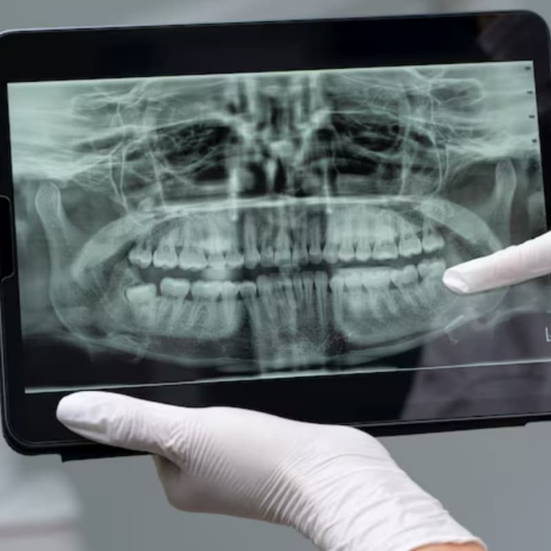
Panoramic X-Ray
Panoramic X-rays provide a broad image of the entire mouth, including the upper and lower jaws, wisdom teeth, jaw joints, and surrounding facial structures. Because this scan offers a wider perspective, it is often used when planning orthodontic treatment, evaluating wisdom teeth, or assessing jaw-related conditions. In some cases, this type of scan may influence how often dental x ray needed, especially when dentists need to monitor changes across the entire jaw over time.
Cone Beam CT (3D Imaging)
Cone Beam CT scans produce highly detailed three-dimensional images of the teeth, jawbone, nerves, and surrounding structures. This advanced imaging technology is typically reserved for complex dental procedures such as dental implant placement, impacted tooth evaluation, or jaw surgery planning. Because it provides extremely precise information, it is usually recommended only when necessary for advanced diagnosis or treatment planning.
Quick Comparison Table
| X-Ray Type | Purpose | Typical Use |
|---|---|---|
| Bitewing | Detect cavities | Routine checkups |
| Periapical | Root and bone diagnosis | Infection detection |
| Panoramic | Full mouth overview | Wisdom teeth analysis |
| CBCT | 3D imaging | Implant planning |
Each imaging method contributes to determining how often dental x ray needed for accurate diagnosis.
Are Dental X-Rays Safe?
One of the biggest concerns patients have is radiation exposure.
Fortunately, modern digital imaging systems use extremely low radiation levels.
Radiation Exposure Comparison
| Source | Radiation Dose |
|---|---|
| Digital Dental X-Ray | ~0.005 mSv |
| Chest X-Ray | ~0.1 mSv |
| Natural daily background radiation | ~0.01 mSv |
Digital dental imaging uses up to 80–90% less radiation than older film-based systems.
Dentists also follow strict safety measures:
- Lead apron protection
- Thyroid collars
- Minimal exposure time
- Imaging only when necessary
Because of these safety measures, understanding how often dental x ray needed should not create unnecessary concern for most patients.
Dental X-Rays and Professional Teeth Cleaning
Dental imaging often works together with routine cleaning and scaling procedures.
During a professional cleaning visit, dentists may recommend imaging to check for hidden plaque buildup, bone loss, or cavities beneath the gumline.
If you’re curious about diagnostic imaging in northern Singapore, this related guide about dental x ray in Yishun explains how modern clinics combine X-ray diagnostics with preventive treatments.
Pro Tip: Combining routine scaling with periodic imaging ensures dentists can monitor oral health both above and below the gumline, which improves long-term outcomes.
Signs You Might Need a Dental X-Ray Sooner
Sometimes imaging is necessary before your next scheduled dental exam.
Warning Signs
You may need an X-ray sooner if you experience:
- Persistent tooth pain
- Swelling around gums
- Loose teeth
- Sudden sensitivity to hot or cold foods
- Difficulty chewing
If any of these symptoms appear, dentists may recommend imaging regardless of your previous schedule to reassess how often dental x ray needed in your situation.
Conclusion: Where to Get Safe and Accurate Dental X-Rays
Understanding how often dental x ray needed helps patients stay proactive about oral health. Imaging allows dentists to detect hidden cavities, monitor bone health, and prevent serious dental problems before they progress.
Modern dental clinics now use advanced digital technology that provides fast, precise images with extremely low radiation exposure. When combined with routine dental checkups, these scans become a powerful preventive tool.
If you’re considering a professional diagnostic evaluation, you can explore the complete procedure and benefits of Dental X-Ray services here to see how modern imaging supports early detection and personalised treatment planning.
By scheduling regular checkups and following your dentist’s recommendations on how often dental x ray needed, you can maintain healthier teeth, avoid complex treatments, and protect your long-term oral health.
Pro Tip: Don’t wait until tooth pain appears. Scheduling routine exams and diagnostic imaging at the right intervals can save both time and costly treatments later on.
FAQs (Frequently Asked Questions)
How often dental x ray needed for healthy adults?
For adults with good oral health and low cavity risk, dentists usually recommend dental X-rays every two to three years. The exact schedule may vary depending on individual risk factors and oral health history. This helps determine how often dental x ray needed to monitor areas that cannot be seen during a regular dental exam.
How often dental x ray needed for children?
Children may need dental X-rays every 6–12 months because their teeth and jaw structures are still developing. Imaging helps dentists monitor tooth growth, detect early cavities, and identify potential alignment issues. This is why dentists carefully evaluate how often dental x ray needed for younger patients.
Can dental X-rays detect cavities early?
Yes, dental X-rays can detect cavities forming between teeth or under existing fillings before they become visible. Early detection allows dentists to treat decay sooner and prevent it from spreading. This is one reason professionals assess how often dental x ray needed for each patient.
Do dental X-rays hurt?
No, dental X-rays are painless and usually take only a few minutes to complete. Patients may feel slight pressure from the sensor, but the procedure is quick and comfortable. Modern technology also uses very low radiation levels, making it safe when determining how often dental x ray needed.
5. Can I skip dental X-rays if my teeth feel fine?
Not always, because many dental problems develop without noticeable symptoms. X-rays allow dentists to detect hidden issues such as early decay, bone loss, or infections. Regular imaging helps determine how often dental x ray needed to maintain long-term oral health.